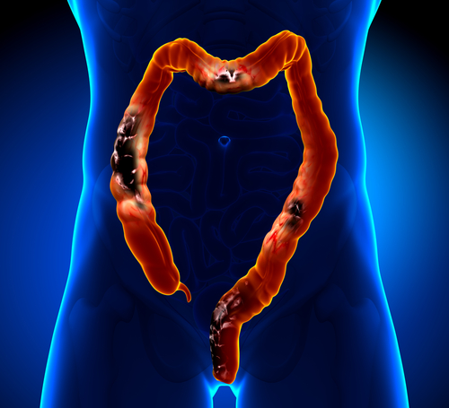
 The American Society for Gastrointestinal Endoscopy (ASGE) and the American Gastroenterological Association (AGA) released a series of recommendations regarding dysplasia associated with inflammatory bowel disease (IBD). The document published in the journal GIE: Gastrointestinal Endoscopy and Gastroenterology aims to improve and standardize the methods of surveillance and management of the condition.
The document is meant to be a statement of consensus about the treatment and diagnosis of dysplasia. Ulcerative colitis and Crohn’s disease, the two most common forms of IBD, are diseases that develop in the region of the colon and increase the risk of suffering from colorectal cancer, which are thought to emerge from dysplasia areas, or abnormal cellular alterations in the colon mucosa.
The diagnosis of the condition is usually made with mucosa examination through the conduction of targeted biopsies in the visible lesions, as well as extensive random biopsies to search for invisible dysplasia. However, the two associations want to modernize procedures, as current endoscopy technologies enable the visibility of most dysplasia, as announced in a press release.
“In the field of gastrointestinal endoscopy, we are fortunate to have new types of equipment and technology that provide high-definition visualization of the colon,” said ASGE President Colleen M. Schmitt, MD, MPH, FASGE. “In addition, gastrointestinal endoscopists are continually updating their skills to stay abreast of the latest techniques for surveillance, as well removal of lesions. These procedures contribute greatly to the evolving ability of the health care team to help patients with IBD achieve optimal health and minimize their risk for colon cancer.”
The new recommendations include nine statements about the detection of lesions, as well as the optimal management of the disease in patients who also suffer from IBD. The statements were designed by a panel of international specialists and stakeholders in the field, based on the Institute of Medicine’s standards of care. However, the group did not reach consensus regarding the use of random biopsies when endoscopists use high-definition white-light colonoscopy or chromoendoscopy.
The updated recommendations are the result of the need to conduct more frequent and better chromoendoscopy procedures in IBD patients, as a way of improving visibility of the tissue.
- “When performing surveillance with white-light colonoscopy, high definition is recommended rather than standard definition.
- When performing surveillance with standard-definition colonoscopy, chromoendoscopy is recommended rather than white-light colonoscopy.
- When performing surveillance with high-definition colonoscopy, chromoendoscopy is suggested rather than white-light colonoscopy.
- When performing surveillance with standard-definition colonoscopy, narrow-band imaging (NBI) is not suggested in place of white-light colonoscopy.
- When performing surveillance with high-definition colonoscopy, narrow-band imaging is not suggested in place of white-light colonoscopy.
- When performing surveillance with image-enhanced high-definition colonoscopy, narrow-band imaging is not suggested in place of chromoendoscopy.
- After complete removal of endoscopically resectable polypoid dysplastic lesions, surveillance colonoscopy is recommended rather than colectomy.
- After complete removal of endoscopically resectable nonpolypoid dysplastic lesions, surveillance colonoscopy is suggested rather than colectomy.
- For patients with endoscopically invisible dysplasia (confirmed by a GI pathologist) referral is suggested to an endoscopist with expertise in IBD surveillance using chromoendoscopy with high-definition colonoscopy.”

“A diagnosis of inflammatory bowel disease is often overwhelming and frightening for patients,” said the president of the AGA Institute, John I. Allen, MD, MBA, AGAF. “These patients not only have the immediate concern of how the disease affects their life, they also face an increased risk of developing colorectal cancer, which needs intense surveillance. We hope these guidelines will help gastroenterologists and their patients develop the most appropriate and effective monitoring system to reduce their risk of developing colorectal cancer.”
The group comprised of endoscopists, pathologists, nurses, IBD experts and patients was led by Loren Laine, MD, AGAF, from the Yale School of Medicine and VA Connecticut Healthcare System, who explained that the group focused not only on management techniques, but also on patients’ preferences, since for instance patients may prefer to undergo a colectomy when the risk of developing colorectal cancer is higher, or to delay it in cases of dysplasia.
“We are now seeing more and more of what used to be considered ‘invisible’ lesions,” said Laine. “Given that, and the fact that we can remove many of the lesions endoscopically, we have a higher comfort level in using surveillance with these patients, rather than more invasive treatment. This is good news for IBD/colitis patients, particularly those who have a significant portion of the colon involved.”
The specialists in the group further noted that there is a lack of resources to train young endoscopists and increase IBD surveillance. They believe that further research would increase knowledge and training regarding chromoendoscopy with high-definition colonoscopy, definition of appropriate surveillance gaps for procedure and natural history of both visible and endoscopically invisible dysplasia.

