
 A new nanoscale agent for imaging the gastrointestinal (GI) tract was developed by a multi-institutional team of researchers. According to the journal Nature Nanotechnology, which recently published the results of a study, the new method is a safe and noninvasive way of assessing the function and properties of the GI tract in real time, which may help physicians perform better diagnosis of diseases such as IBD, as well as improve gut disease treatments.
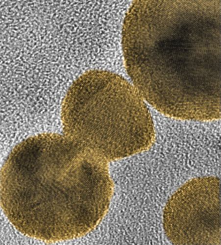
The scientists designed a multimodal functional imaging agent through a complementary approach that uses both photoacoustic imaging and positron emission tomography (PET). The research team expects to use the agent to perform noninvasive functional imaging of the intestine in real time. They developed a family of nanoparticles that are able to provide good optical contrast for imaging and avoid absorption into the body, as well as withstand the harsh conditions of the stomach and intestine.
Weibo Cai, associate professor of radiology, medical physics and biomedical engineering at the University of Wisconsin-Madison, worked in collaboration with Jonathan Lovell, assistant professor of biomedical engineering at the State University of New York at Buffalo, and Chulhong Kim, assistant professor of creative information technology engineering at Pohang University of Science and Technology in South Korea in order to develop the agent.
The team performed clinical trials in mice and is now planning to conduct new ones with human population. “This is one of the first studies using both imaging techniques,” Cai explained. “The two imaging techniques work well together and get us all of the information that we need.” Lovell is also excited about the project’s outcomes for patients, as he stated that they “could potentially induce a paradigm shift that allows for much more routine examination of the intestine function, that would really benefit overall health.”
Small bowel bacterial overgrowth, irritable bowel syndrome, and inflammatory bowel disease are examples of diseases that occur in the intestine and that can cause serious complications. Physicians aren’t currently able to functionally image the intestine, and until now it was common to perform X-rays and ultrasound on patients after they drank a chalky liquid called barium and technicians. However, there were still many limitations, as accessibility and the possibility of radiation exposure.
With this new method, patients will also drink a liquid containing nanoparticles with bright dyes in order to allow an imaging technician to noninvasively examine the illuminated intestine with photoacoustic imaging. “We can actually see the movement of the intestine in real time,” Lovell said.
The collaboration of the researchers is what enabled the discovery, since Cai is specialized in PET imaging, which uses radioisotope-based tracers and is used in health care settings for noninvasive, whole-body imaging. Both Lovell and Kim are experts in photo acoustic imaging, a technique that draws on ultrasound to generate high-definition images through light-based imaging. PET imaging can penetrate deeper and image the entire body, on contrary to photoaccoustic techniques.
Cai said that the purpose of the team is that the agent can be used in the diagnosis of certain disease-related markers, as well within therapeutic applications in the near future. “It is everything I would hope for in an imaging agent, and it is safe since we use FDA-approved agents to make these nanoparticles. That is why I am so excited about this, these are the promising first steps,” he said.